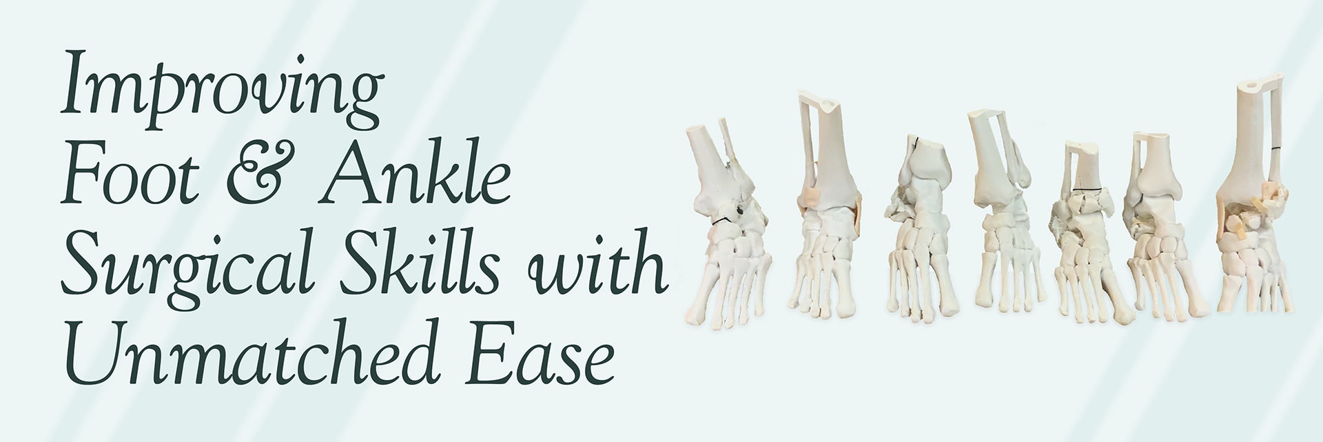
Improving Foot and Ankle Surgical Skills with Unmatched Ease
Surpassing cadaveric training with custom surgical models that prepare surgeons for complex cases.
With traditional cadaver labs, complex surgical cases are extremely difficult to train. Obtaining a cadaver with just one specific deformity for practice can be a logistical nightmare. It would be impossible to provide a cadaver with multiple deformities and pathologies. Additionally, cadaveric training comes with a cost, both an ethical and cost perspective.
The lack of readily available orthopedic surgeon training in complicated foot and ankle surgery ultimately hurts podiatric surgeons and their patients. It denies them the discovery of surgical tools and products that make repairing injuries easier and more successful.
Our customer wanted to remedy this by showing their products in an advanced surgeon training lab for ankle pathologies and asked us to build custom anatomical models. Since we're always eager to help advance surgical education for better patient outcomes, we were happy to take on this project.
A Challenge to Overcome
Our customer's advanced training lab required a variety of pathologies and deformities including high-energy fracture patterns, avascular necrotic bone, misaligned ankle fusions, and Weber fractures. In light of cadaver limitations, they couldn't meet these requirements with traditional cadaveric training.
A Much-Needed Solution
Based on the customer's specifications, we created 7 patient-specific foot and ankle STUDs. There were a number of cases submitted by the course faculty that presented complex situations in which our customer's products could be utilized. After helping our customer choose the most impactful cases, we modeled and manufactured relevant ankle models.
Driving Results That Make an Impact
The anatomical foot models were a valuable tool for educating attendees on how to best serve patients, enabling them to develop informed and well-rounded approaches to surgical procedures. Participants reviewed patient histories and CT scans before seeing and feeling the actual bones, which provided them with a comprehensive understanding of the specific anatomy and pathology involved in the complex cases. By having a physical representation of the anatomy in front of them, attendees could visually grasp the intricacies of the patient's condition, facilitating discussions on the most effective plan of action. The models were a valuable tool for educating attendees on how to best serve patients, enabling them to develop informed and well-rounded approaches to surgical procedures.
Do you need a custom solution for a complex training problem? Get in touch with us today to find out how we can help!


